T wave normally becomes inverted during the first week of life. The QRS complex is the combination of three of the graphical deflections seen on a typical electrocardiogram ECG or EKGIt is usually the central and most visually obvious part of the tracing.
Right Bundle Branch Block Rbbb Litfl Ecg Library Diagnosis
Normally when the QRS is narrow there should be predominant negativity in lead V1.
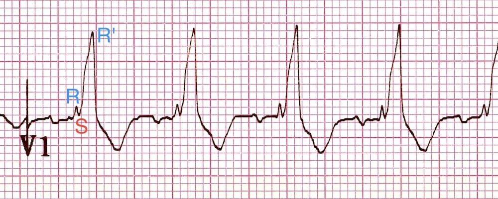
. A monophasic R or biphasic qR complex in V1 An RSR or bunny ear pattern present in V1 or V2 with the R peak higher in amplitude than the R peak see image below A rS complex in lead. The only break from this pattern of progression are foci originating from the AMC which have classically have a qR in V 1 17 which may be due to a unique exit from remnant His-Purkinje tissue. The expected ST-segmentT-wave configurations are discordant directed opposite from the terminal portion of the QRS complex.
Typical RSR pattern M-shaped QRS in. Never connect pedals via USB and RJ12 at the same time. Tables 1 and 2.
T wave is normal. Upright T wave in V1 1 week of age. RS ratio in V1 and V2 ULN for ageor RS ratio in V6 1 after 1 month of age Upright T in V1 older than 3 days provided that the T is upright in V5 and V6.
Newborns have upright T wave in V1. Pediatric EKG Interpretation. 左脚ブロック v1で qs 型かつ v6 で rr rsr 型 c.
The more anterior RVOT free wall will transition later V 4 or V 5 than the septal RVOT V 3 or V 4 which is more posterior Figure 2. In adults the QRS complex normally lasts 80 to. Criteria for IRBBB Incomplete Right Bundle Branch Block are satisfied because in addition to the qR pattern in lead V1 there are terminal S waves in lateral leads I and V6.
Abnormal depolarization of both ventricles also causes abnormal repolarization. RSR in V1 with a tall R 10 mm qR pattern in V1. 左脚前枝ブロック lad -45 -90度qrs が 009 010秒 pq 軽度延長i に異常 q波 qr型 iiiii で rs型 s は iii の方が深く大きい.
Upright T wave beyond the first week of life is a strong indicator of RVH. The paced QRS morphology during unipolar LBBP shows the pattern of right bundle branch block RBBB in V1 lead qR or Qr or improving the LBB conduction in patients with LBBB Figure 3C 31 32. Before you read the EKG look for.
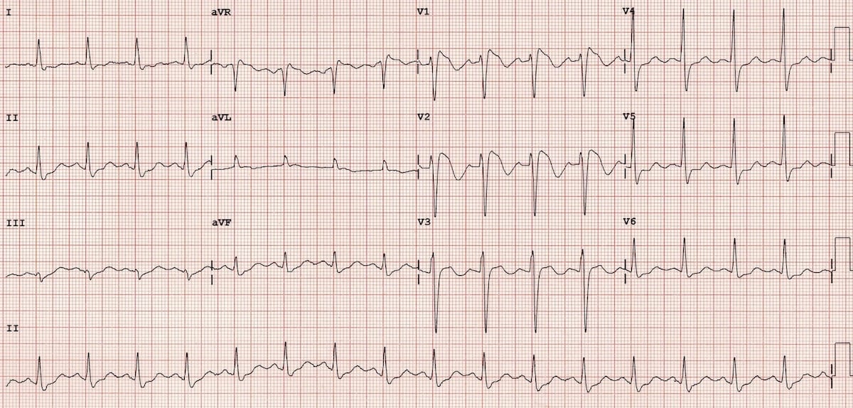
Wide QRS complex 120 ms. An rsr with widening of the qrs and characteristic findings in other leads is due to a right bundle branch block. Lead V1 shows an rS pattern and lead V6 shows a qR pattern.
The QRS complex in leads V1 to V3 may assume any of the following morphologies. Sometimes rather than an RSR pattern in V1 there may be a broad monophasic R wave or a qR complex. This pattern is called QRS-complexT-wave axis.
A qR pattern in V1 suggests RVH make sure there is not a small r in an rsR configuration. A monophasic R or biphasic qR complex in V1 A rS complex in lead V6 favors VT A RSR or bunny-ear pattern present in V1 with the R peak being higher in. This produces a saddle-shaped ST segment that the computer may mistake for acute ischemia.
The RBBB pattern is usually incomplete and is influenced by the level of capture of the distal His bundle or proximal left bundle distal. There is a qR pattern in lead V1 which is distinctly abnormal. Rsr pronounced r s r-prime can be a normal finding in leads v1 and v2.
Jun 17 2019 Longitudinal multicenter retrospective cohort study from the eICU Collaborative Research Database V1. Lead V1 shows an rSR pattern broad R wave and lead V6 shows a qRS pattern broad S wave. Shorty URL Permanent shortened hyperlink to content status page.
Improved smoothness of the load cell brake pedal signal. In case you want to compare old and new or revert to the old one - change Firmware Manager to manual update open the Updater and then use the 3 dots to select version. However papillary muscle PVCs lack the typical rsR in V1 and Q waves in limb leads have a less sharp QRS onset an overall longer QRS duration and pleomorphic PVC morphologies due to multiple exit sites Table 22425 PVCs originating from moderator band or false tendons can have two disparate morphologies or axes as the ectopy exits from.
Site Alerts Tracking and change info preserved and made. Redux state change causes unwanted re render when using custom route react-router v6. Pedals need to be plugged in via USB for the firmware update.
Electrocardiogram with right bundle branch block. Full standard is two large squares 1 mV 10 mm and half standard is one large square 05mV 5 mm. Patient age as many values change with age.
T wave remains inverted in V1 throughout early childhood. V1で rsr 型かつ v6 に幅広いs b. It corresponds to the depolarization of the right and left ventricles of the heart and contraction of the large ventricular muscles.
An upright T in V1 is not abnormal in patients 6 years or older. In some cases the rSr or qR pattern may combine with a mild degree of benign anterior ST segment elevation aka male pattern.

The Rsr Pattern In Leads V1 V2 Algorithm And Differential Diagnosis Sciencedirect

Right Bundle Branch Block Rbbb Litfl Ecg Library Diagnosis

Rsr In V1 Resources

Differential Diagnosis Of Rsr Pattern In Leads V1 V2 Comprehensive Review And Proposed Algorithm Baranchuk 2015 Annals Of Noninvasive Electrocardiology Wiley Online Library

The Rsr Pattern In Leads V1 V2 Algorithm And Differential Diagnosis Sciencedirect

Dr Smith S Ecg Blog Rsr With St Elevation Is This Right Bundle Branch Block With Stemi Type 2 Brugada

Dr Smith S Ecg Blog Rsr With St Elevation Is This Right Bundle Branch Block With Stemi Type 2 Brugada
Right Bundle Branch Block Rbbb Litfl Ecg Library Diagnosis
0 comments
Post a Comment